Lipid Expansion Microscopy
Abstract
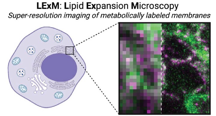
Strategies to visualize cellular membranes with light microscopy are restricted by the diffraction limit of light, which far exceeds the dimensions of lipid bilayers. Here, we describe a method for super-resolution imaging of metabolically labeled phospholipids within cellular membranes. Guided by the principles of expansion microscopy, we develop an all-small molecule approach that enables direct chemical anchoring of bioorthogonally labeled phospholipids into a hydrogel network and is capable of super-resolution imaging of cellular membranes. We apply this method, termed lipid expansion microscopy (LExM), to visualize organelle membranes with precision, including a unique class of membrane-bound structures known as nuclear invaginations. Compatible with standard confocal microscopes, LExM will be widely applicable for super-resolution imaging of phospholipids and cellular membranes in numerous physiological contexts.